Catherine Wagner. Seeing Through Matter

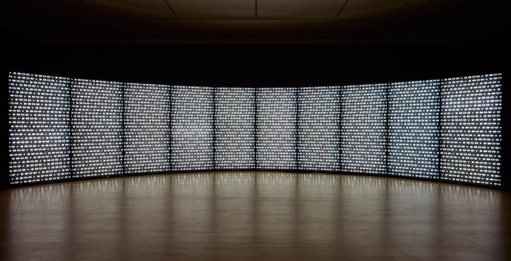
Catherine Wagner began exploring the relationship between science and photography in the early 1990s, photographing petri dishes of algae, clone libraries in deep freezers, and tools like beakers and test tubes in scientific labs. In 1997 the San José Museum of Art awarded her the two-year Artist Residency Fellowship to continue her investigation inside Silicon Valley laboratories. Rather than photograph science from the surface, Wagner used a medical imaging device—magnetic resonance imaging (MRI)—in lieu of her camera. “I wondered what it might look like if I was to literally see through matter,” said the artist.1 Her first imaging experiments in the MRI labs at Stanford University were with pumpkins, pomegranates, and onions. Wagner took the digital information and produced photographic images with the cross-sectioned views of their interiors. For Pomegranate Wall (2000), the artist used such images of the pomegranate, which were particularly striking for their resemblance to human cells, repeating them in a pattern throughout a forty-foot arc of backlit images and evoking the organic process of cellular replication on a grand scale.
Catherine Wagner, acknowledgments for Catherine Wagner: Cross Sections (San José, CA: San José Museum of Art; and Santa Fe, NM: Twin Palms Publishers, 2001), n.p. ↩︎